Detoxcication of Benzophenanthridine alkaloids
Detoxicitation of Benzophenanthridine alkaloids
Saguinarine reductase drives a detoxifying cycle
The ability of plants to protect their cells from adverse effects of self-made toxins
is an important though less understood determinant of the evolutionary success of secondary biosynthesis.
Benzophenthridines display a variety of cytotoxic effects, that make them highly effective phytoalexins: intercalation into dsDNA,
inhibition of membrane potential dependent enzymes, binding to the cytoskeleton, etc.
We could show that the benzophenanthridines do not accumulate inside living cells of Eschscholzia, but are excreted into the
apoplast and external medium. External alkaloids, preferably sanguinarine, undergo a rapid re-uptake which is tíghtly coupled
to their intracellular reduction. The less toxic dihydro-alkaloids are further derivatized according to the biosynthetic route, thus
forming less toxic members of the benzophenanthridine group. The cycle allows to present the phytoalexin sanguinarine at the
cellular surface without taking the risk to intoxicate cells by critical levels of their own toxin.

Excretion of benzophenanthridines into the apoplast area.
Celles were loaded with a fluorescein dye that accumulatess in intact vacuoles (green) thus indicating cell viability.
Red colour symbolizes benzophenanthridine fluorescence (proven in previous experiments by local fluorescence spectra).
left: typical cell string at the time of adding yeast elicitor.
middle: a similar cell string after 20 h elicitor treatment
right: same cell string as in left image, after 20 h elicitor treatment. For better visualization of the cell wall region, cells were plasmolyzed prior to photography.
Some vacuolar (v), cytoplasmic (cyt) and cell wall (cw) regions are marked.
Note that the alkaloid fluorescence sticks to the apoplast even after plasmolysis.
(From Weiss et al. 2006)
|

Uptake and conversion of sanguinarine by intact cells of E. californica.
Cultured cells were incubated with 10 µM sanguinarine.
This alkaloid shows here a red flourescence, dihydro-sanguinarine shows blue fluorescence.
Fluorescence images were taken after 1 min (left), 12 min (middle) and 30 min (right).
Some vacuolar (v), cytoplasmic (cyt) and cell wall (cw) regions are marked.
(From Weiss et al. 2006).
|

|
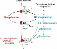
Schematic view of the recycling of
benzophenanthridine alkaloids by intact cells of Eschscholzia.
Sanguinarie reductase (SR) is a soluble protein of 29.5 kDa. The isolated enzyme catalyses the
NAD(P)H-dependent reduction of sanguinarine with a Km (Sang) of about 10 µM.
The transport steps around the reduction of sanguinarine and the oxidation of dihydroalkaloids are unknown and under work.
|
Zum Seitenanfang


